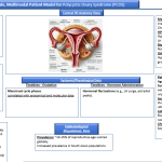
Patient Model - Molecular, Cellular, Tissue, Organ, Systemic, Clinical scales


A 32-year-old woman presents with irregular periods for the past year, severe hot flashes, and difficulty conceiving for 2 years.
- History of autoimmune thyroiditis.
- No significant gynecological surgeries or infections.
Key Diagnosis: Determines ovarian reserve and function.
Hormonal Assays:
a) Anti-Müllerian Hormone (AMH): Reflects ovarian reserve (low levels indicate diminished reserve).
b) Follicle-Stimulating Hormone (FSH): Elevated levels suggest poor ovarian function.
c) Estradiol (E2): Assessed on day 2-5 of the menstrual cycle; elevated levels can indicate reduced ovarian reserve.
Ultrasound Imaging:
Antral Follicle Count (AFC): Transvaginal ultrasound counts small follicles (2-10 mm) in both ovaries; fewer follicles suggest diminished reserve.
Ovarian Volume Assessment:
Done using transvaginal ultrasound; reduced ovarian volume may indicate follicle depletion.
Dynamic Testing:
Clomiphene Citrate Challenge Test (CCCT): Evaluates ovarian response by measuring FSH levels before and after administering clomiphene citrate.
Molecular Level:
- AMH: 0.2 ng/mL (Low, indicating diminished ovarian reserve).
- Estradiol: 95 pg/mL (Elevated due to compromised ovarian function).
Cellular Level:
- Ovarian granulosa cell apoptosis rate: Increased in follicle biopsy.
Tissue Level:
- Antral Follicle Count (AFC): 3 (Normal range: 6-10 in this age group).
- Ovarian stromal volume reduced on transvaginal ultrasound.
Organ Level:
- Ovarian volume: 2 cm³ (Normal: >4 cm³).
Systemic Level:
- FSH: 18 mIU/mL (Elevated; normal: <10 mIU/mL).
- LH: 12 mIU/mL (Elevated; normal: <10 mIU/mL).
Epidemiological Level:
Incidence of premature ovarian insufficiency in women under 40: ~1%.


A 28-year-old woman with secondary infertility after a D&C procedure for a missed miscarriage.
- Complains of light menstrual flow and dysmenorrhea.
- History of recurrent intrauterine infections.
Key Diagnosis: Determines ovarian reserve and function.
Hysteroscopy:
- Gold standard for directly visualizing and assessing intrauterine adhesions and scarring.
Sonohysterography (Saline Infusion Sonography):
- Uses saline infusion during transvaginal ultrasound to identify intrauterine adhesions or irregularities.
Hysterosalpingography (HSG):
- X-ray with contrast to outline the uterine cavity; detects structural abnormalities and adhesions.
MRI of the Uterus:
- Provides detailed imaging for more severe cases of uterine scarring or for preoperative planning.
Endometrial Biopsy:
Evaluates endometrial thickness and health for functional assessment.
Molecular Level:
- Transforming growth factor-beta (TGF-β): Elevated, associated with fibrosis.
- Vascular endothelial growth factor (VEGF): Reduced in endometrial biopsy.
Cellular Level:
- Fibroblast proliferation rate: Elevated in scar tissue.
Tissue Level:
- Endometrial thickness: 4 mm on sonohysterography (Normal: >7 mm during the proliferative phase).
- Presence of dense intrauterine adhesions on hysteroscopy.
Organ Level:
- Uterine cavity distorted with synechiae.
Systemic Level:
- Hormone levels (FSH, LH): Normal; absence of systemic endocrine dysfunction.
Epidemiological Level:
Prevalence of Asherman’s Syndrome after D&C: ~1.5%.


A 35-year-old man with azoospermia diagnosed during fertility evaluation.
- Reports reduced libido and fatigue for 1 year.
- History of mumps orchitis during adolescence.
Key Diagnosis: Determines the presence and functionality of spermatogenic cells.
Hormonal Assays:
- Testosterone: Low levels may indicate hypogonadism.
- FSH and LH: Elevated FSH levels suggest primary testicular failure.
Semen Analysis:
- Evaluates sperm count, motility, and morphology. Absence of sperm suggests azoospermia.
Testicular Ultrasound:
- Assesses testicular volume, structure, and the presence of abnormalities such as varicocele or microcalcifications.
Testicular Biopsy:
- Detects the presence of spermatogenic cells and evaluates the degree of spermatogenesis.
Genetic Testing:
Identifies chromosomal abnormalities (e.g., Klinefelter syndrome, Y-chromosome microdeletions) causing testicular failure.
Molecular Level:
- Testosterone: 2.1 ng/mL (Low; normal: >3.5 ng/mL).
- FSH: 24 mIU/mL (High; normal: 1-10 mIU/mL).
- LH: 12 mIU/mL (Elevated; normal: 1-8 mIU/mL).
Cellular Level:
- Sertoli cell degeneration noted on testicular biopsy.
- Germ cell apoptosis rates elevated.
Tissue Level:
- Loss of spermatogenic cells in seminiferous tubules.
- Thickened basement membrane of seminiferous tubules.
Organ Level:
- Testicular volume: 8 mL (Normal: 15-20 mL).
Systemic Level:
- Decreased sperm count in semen analysis: 0 sperm (azoospermia).
Epidemiological Level:
Prevalence of non-obstructive azoospermia: ~10-15% of infertile men.


A 30-year-old woman with recurrent implantation failure despite three cycles of IVF.
- Complains of heavy, irregular periods and pelvic pain.
- No significant infections or surgeries.
Key Diagnosis: Determines ovarian reserve and function.
Endometrial Thickness Measurement:
- Assessed using transvaginal ultrasound; thin endometrium (<7 mm) is associated with poor implantation.
Sonohysterography:
- Highlights abnormalities in the endometrial cavity, such as polyps, fibroids, or scarring.
Endometrial Biopsy:
- Provides histological evaluation for chronic inflammation, infection, or insufficient development.
Doppler Ultrasound:
Assesses blood flow to the endometrium, which correlates with endometrial receptivity.
Molecular Level:
- Pro-inflammatory cytokines (IL-6, TNF-alpha): Elevated in endometrial biopsy.
- Integrin αvβ3 (implantation marker): Decreased expression.
Cellular Level:
- Endometrial epithelial cell density: Reduced.
Tissue Level:
- Endometrial thickness: 5 mm on Day 10 of the cycle (Normal: 8-14 mm).
Organ Level:
- Uterine artery blood flow: Resistance Index (RI) of 0.8 (Normal: <0.6).
Systemic Level:
- Hormone levels (FSH, LH, E2): Normal.
Epidemiological Level:
Prevalence of thin endometrium in recurrent implantation failure: ~10-20%.


A 25-year-old woman with ovarian insufficiency after chemotherapy for Hodgkin’s lymphoma.
- Reports amenorrhea for 1 year post-treatment.
- No prior fertility preservation.
Key Diagnosis: Evaluates the extent of gonadal damage after chemotherapy or radiotherapy.
AMH and FSH Levels:
- Assess ovarian reserve and function in women post-treatment.
Antral Follicle Count (AFC):
- Quantifies the ovarian reserve using transvaginal ultrasound.
Testicular Biopsy:
- Determines residual spermatogenesis in men.
MRI or CT Scans:
Evaluate pelvic or testicular anatomy for structural damage.
Molecular Level:
- AMH: 0.1 ng/mL (Severely low).
- Reactive oxygen species (ROS): Elevated in ovarian tissue.
Cellular Level:
- Follicular atresia and increased apoptosis in ovarian biopsy.
Tissue Level:
- Antral Follicle Count (AFC): 0.
- Ovarian stromal fibrosis visible on ultrasound.
Organ Level:
- Ovarian volume: <2 cm³ (Significantly reduced).
Systemic Level:
- FSH: 40 mIU/mL (High; indicative of ovarian failure).
Epidemiological Level:
Incidence of premature ovarian insufficiency post-chemotherapy: ~30%.


Name: Mrs. A, 29 years old
Chief Complaint: Recurrent pregnancy loss and difficulty conceiving for 2 years.
History:
- Regular menstrual cycles with normal flow.
- No significant gynecological infections or surgeries.
- Underwent hysterosalpingography (HSG), which suggested a uterine anomaly.
Suspected Diagnosis: Uterine Septum (Congenital Müllerian Anomaly)
Key Diagnosis: Detects congenital or acquired structural abnormalities that impair function.
3D Transvaginal Ultrasound:
- Identifies uterine anomalies, such as septa or bicornuate uterus.
MRI:
Provides detailed imaging of reproductive anatomy for surgical planning.
Molecular Level:
- HOXA10 & HOXA11 gene expression: Reduced (indicative of impaired uterine receptivity).
- VEGF (Vascular Endothelial Growth Factor): Decreased (impaired endometrial blood flow).
Cellular Level:
- Endometrial cell proliferation rate: Lower in septal region (deficient response to hormonal signaling).
- Fibrotic markers (TGF-β, Collagen Type I): Increased in septal tissue.
Tissue Level:
- Endometrial thickness: 6 mm in septal region (Normal range: 8-12 mm).
- Histopathology of Septal Tissue: Poor glandular development, reduced vascularization.
Organ Level:
- 3D Transvaginal Ultrasound Findings: Uterine septum measuring 12 mm (Normal: No septum).
- MRI Findings: Fundal indentation >15 mm (consistent with septate uterus).
Systemic Level:
- Hormonal Profile: Normal estrogen and progesterone levels.
- Menstrual Cycle Regularity: Normal but suspected poor implantation due to septum.
Epidemiological Level:
Associated Risk: Septate uterus linked to a 44% miscarriage rate without surgical correction.
Prevalence of Uterine Septum: ~3-7% of women in the general population.


Name: Mr. B, 34 years old
Chief Complaint: Infertility for 3 years, confirmed non-obstructive azoospermia.
History:Normal libido and secondary sexual characteristics.
No history of infections or surgeries.
Genetic evaluation pending.
Suspected Diagnosis: Y-Chromosome Microdeletion (Yq11 AZF Region Deletion)
Advanced Techniques:
Evaluates cellular integrity and pathological changes in ovarian, uterine, or testicular tissues.
Proteomics and Genomics:
Analyze molecular markers of tissue damage or infertility.
Flow Cytometry:
Quantifies specific cell types, such as stem cells or spermatogenic cells.
Tissue Staining and Histology:
Molecular Level:
- Sperm DNA Fragmentation Index (DFI): 48% (Severe DNA damage; normal: <15%).
- AZFa, AZFb, AZFc Gene Deletions: Present in AZFb region.
- Testosterone Biosynthesis Genes (CYP17A1, HSD17B3): Normal.
Cellular Level:
- Germ Cell Density in Testicular Biopsy: Severely reduced.
- Sertoli Cell-Only Syndrome: Confirmed on histology.
Tissue Level:
- Spermatogenesis: Completely absent in seminiferous tubules.
- Testicular Fibrosis: Moderate interstitial fibrosis observed.
Organ Level:
- Testicular Volume (Ultrasound): 9 mL (Normal: 15-25 mL).
- Scrotal Doppler Study: Normal vascular supply, no varicocele.
Systemic Level:
- FSH: 22 mIU/mL (Elevated; normal: 1-10 mIU/mL).
- LH: 11 mIU/mL (Elevated; normal: 1-8 mIU/mL).
- Testosterone: 4.2 ng/mL (Normal but at the lower range).
Epidemiological Level:
Chance of Sperm Retrieval in AZFb Deletion: <5% with TESE (Testicular Sperm Extraction).
Prevalence of Y-Chromosome Microdeletion in Azoospermic Men: ~10-15%.